


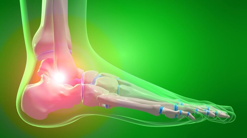
Osteoarthritis of the ankle joint is a chronic disease that affects the articular cartilage and then other structures of the joint (capsule, joint, bones, ligaments). It has a degenerative-dystrophic nature. It manifests as pain and limited movement, followed by a gradual impairment of support and walking functions. Diagnosis is based on symptoms, examination and radiography. Treatment is usually conservative, anti-inflammatory drugs, chondroprotectors and glucocorticoids are used, and exercise therapy and physical therapy are prescribed. In severe cases, medical arthroscopy, arthrodesis or endoprosthesis is performed.
general information
Osteoarthritis of the ankle joint is a disease in which the articular cartilage and the surrounding tissues are gradually destroyed. The disease is based on degenerative-dystrophic processes, arthritis is secondary. Arthrosis has a chronic wave-like course, with alternating remissions and exacerbations, and progresses gradually. Women and men suffer equally often. The probability of development increases sharply with age. At the same time, experts note that the disease is "getting younger" - currently every third ankle arthrosis is detected in people under the age of 45.
Cause
Primary arthrosis occurs for no apparent reason. Secondary damage to the ankle joint develops as a result of some unfavorable factors. In both cases, the basis is a violation of the metabolic processes of cartilage tissue. The main causes and predisposing factors for the development of secondary arthrosis of the ankle joint are as follows:
- severe intra- and periarticular injuries (talus bone fractures, ankle fractures, ligament tears and ligament tears);
- ankle surgery;
- excessive load: too intense sports, long walks or constant standing due to working conditions;
- wearing heels, being overweight, permanent microtraumas;
- diseases and conditions related to metabolic disorders (diabetes, gout, pseudogout, estrogen deficiency in postmenopause);
- rheumatic diseases (SLE, rheumatoid arthritis);
- osteochondrosis of the lumbar spine, intervertebral hernia and other conditions accompanied by pinching of nerves and disruption of the muscular system of the legs and feet.
Less often, the cause of arthrosis is non-specific purulent arthritis, arthritis caused by specific infections (tuberculosis, syphilis) and congenital malformations. Unfavorable environmental conditions and hereditary predisposition play a certain role in the development of arthrosis.
Pathogenesis
Normally, the articular surfaces are smooth, flexible, slide smoothly relative to each other during movement and provide effective shock absorption under load. As a result of mechanical damage (trauma) or metabolic disorders, the cartilage loses its smoothness, becomes rough and inelastic. During movement, the cartilages "rub" and damage each other, which leads to worsening pathological changes.
Due to insufficient amortization, the excessive load is transferred to the underlying bone structure, and degenerative-dystrophic disorders develop in it: the bone is deformed and grows along the edges of the joint area. Due to the secondary trauma and disruption of the normal biomechanics of the joint, not only the cartilage and bone, but also the surrounding tissues suffer.
The joint capsule and the synovial membrane thicken, fibrous degeneration foci form in the ligaments and periarticular muscles. The joint's ability to participate in movements and withstand loads decreases. Instability occurs and pain progresses. In severe cases, the joint surfaces are destroyed, the supporting function of the limb is impaired, and movements become impossible.
Symptoms
Initially, after a significant load, rapid fatigue and slight pain in the ankle joint can be noticed. After that, the pain syndrome becomes more intense, its nature and time of occurrence change. Distinctive features of arthritic pain are as follows:
- Incipient pain. It appears after rest and gradually disappears with movement.
- Dependence on load. Increased pain occurs during exercise (standing, walking) and rapid fatigue of the joint.
- Pain at night. They usually appear in the morning.
The condition changes in waves, during exacerbations the symptoms are more pronounced, in the remission phase they first disappear and then become less intense. The gradual progression of symptoms occurs over several years or decades. In addition to pain, the following manifestations are determined:
- Crackling, squeaking or clicking sounds may occur during movement.
- During an exacerbation, the periarticular area sometimes becomes swollen and reddened.
- Due to the instability of the joint, the patient often twists the leg, causing sprains and ligament tears.
- Stiffness and limitation of movements can be observed.
Complications
During exacerbation, reactive synovitis may occur, accompanied by fluid accumulation in the joint. In later stages, pronounced deformation is revealed. Movements are sharply limited and contractures develop. Support becomes difficult, when moving patients are forced to use crutches or a stick. A reduction or loss of working capacity occurs.
Diagnostics
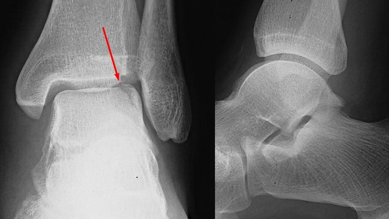
The diagnosis of arthrosis of the ankle joint is made by an orthopedic doctor based on the results of a survey, external examination data and further tests. When examined in the initial stage, changes may not occur, but deformations, movement restrictions, and palpable pain are revealed later. Special importance is attached to visualization techniques:
- X-ray of the ankle joint. It plays a decisive role in establishing the diagnosis and determining the degree of arthrosis. The pathology is indicated by the narrowing of the joint space and the proliferation of the edges of the joint surfaces (osteophytes). At a later stage, cystic formations and osteosclerosis of the subchondral (located under the cartilage) zone of the bone are detected.
- Tomographic examinations. Used when indicated. In difficult cases, in order to more accurately assess the condition of the bone structures, the patient is sent for a computerized tomography examination, and for examination of soft tissues, the patient is sent for an MRI of the ankle joint.
Laboratory tests are unchanged. If necessary, consultation with specialists related to establishing the cause of arthrosis and differential diagnosis with other diseases is prescribed: neurologist, rheumatologist, endocrinologist.
Treatment of ankle arthrosis
Treatment of pathology is long-term and complex. Patients are usually treated by an orthopedic surgeon on an outpatient basis. In the period of exacerbation, hospitalization in the traumatology and orthopedic department is possible. The most important role in slowing the progression of arthrosis is played by lifestyle and appropriate physical activity, therefore the patient receives recommendations for weight reduction and optimizing the load on the leg.
Drug therapy
It is selected individually, taking into account the stage of arthrosis, the severity of symptoms and concomitant diseases. It includes general and local agents. The following drug groups are used:
- Generic NSAIDs. Tablet forms are usually used. Medicines have a negative effect on the mucous membrane of the stomach, so "gentle" medicines should be preferred for gastrointestinal diseases.
- Topical NSAIDs. It is recommended both during the exacerbation period and during the remission phase. It can also be prescribed as an alternative solution if the side effects of the tablets occur. It is available in the form of ointments and gels.
- Chondroprotectors. Substances that help normalize metabolic processes in cartilage tissue. It is used in the form of creams, gels and preparations for intra-articular administration. Use medications containing glucosamine and collagen hydrolyzate.
- Hormonal drugs. In case of severe pain that cannot be relieved by medication, intra-articular corticosteroids are administered up to 4 times a year.
- Metabolic stimulants. In order to improve local blood circulation and activate tissue metabolism, nicotinic acid is prescribed.
Physiotherapy treatment
The patient is prescribed a physical therapy complex, developed taking into account the manifestations and stage of the disease. The patient is referred for physiotherapy. In the treatment of arthrosis, massage and UHF are used. In addition, the following are used in the treatment of pathology:
- laser therapy;
- thermal processes;
- medical electrophoresis and ultraphonophoresis.
Surgery
It is indicated in the later stages of the disease, when conservative therapy is ineffective, severe pain syndrome, deterioration of the patients' quality of life or limited work capacity. The operations are performed in a hospital setting, are open and minimally invasive:
- Arthroscopic interventions. In case of significant damage to the cartilage, arthroscopic chondroplasty is performed. Hygienic arthroscopy (removal of formations that impede movement) is usually performed in case of severe pain occurring in the 2nd stage of arthrosis. The effect lasts for several years.
- Arthrodesis of the ankle joint. It is performed in case of significant damage to the joint surfaces, removing the joint and "fusion" of the bones of the foot and lower leg. It ensures the restoration of the supporting function of the limb in case of loss of joint mobility.
- Endoprosthesis of the ankle joint. It is performed in case of advanced arthrosis. It includes the removal and replacement of the damaged joint surfaces of the bones with plastic, ceramic or metal prostheses. The movements are fully restored, the lifespan of the prosthesis is 20-25 years.
Forecast
The changes that occur in the joint are irreversible, but the slow progression of arthrosis, the timely start of treatment and compliance with the recommendations of the orthopedic traumatologist in most cases make it possible to preserve the ability to work and a high quality of life for decades after the onset. about the first symptoms. With the rapid growth of pathological changes, endoprosthesis makes it possible to avoid disability.
Prevention
Preventive measures include reducing the number of injuries, especially during icy periods in winter. If you are obese, measures should be taken to reduce body weight to reduce the load on the joint. Adhere to the system of moderate physical activity, avoid overloading and microtraumas, and immediately treat diseases that can trigger the development of arthrosis of the ankle joint.