


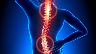
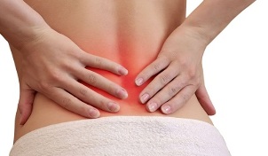
Excessive tension in the back muscles causes a lot of discomfort and pain. Osteopondrosis, which causes damage to the structure of the vertebrae and intervertebral discs, leads to severe stinging of nerve endings. Often, the pathology is accompanied by a deterioration in blood circulation, which provokes disorders in the nutrition of the brain and internal organs.
Osteochondrosis - what is it?

Osteochondrosis is a recurrent type of disease that occurs in chronic form and involves the destruction of vertebrae with intervertebral discs. Their tissues are disturbed, which provokes a decrease in their degree of elasticity with subsequent deformation. The intervertebral space gradually decreases. This causes a loss of spinal stability in areas of pathology development.
The processes of abnormal tissue destruction occur in the background of pinching of nerve endings directed from the site of the spinal cord. As a result, the back muscles are under constant tension. In such a situation, patients complain of back pain and other symptoms.
Based on the peculiarities of the localization of spinal structures covered by degenerative changes, we distinguish between cervical, thoracic, and lumbosacral types of the pathological process. The main symptom of the development of osteochondrosis is pain, the intensity and severity of which usually increase during exercise.
There is also stiffness in movement. In addition, the clinical picture is characterized by the presence of signs of the vertebral type - headache, change in blood pressure, deterioration of visual function, hearing, etc.
Development Mechanism
The development of osteochondrosis is related to the fact that the nucleus pulposus begins to lose its hydrophilic properties. This semi-liquid structure contains connective tissue fibers and chondroitin, a gelatinous material. In the process of development and growth of the human body, the processes of vascular lumbar reduction in the intervertebral plates are actively progressing. Nutrients are supplied in a diffuse manner, which manifests itself in the spontaneous stabilization of concentration. This function becomes the cause of difficulties in the complete restoration of cartilage that has suffered damage or excessive pressure on the spine.
Pathological disorders are more prominent due to violations of hormonal background and human nutrition. Cartilage begins to lack the nutrients needed for normal development. Therefore, the anomalies appear as
- decrease in strength and flexibility;
- changes consistency parameters and configuration properties.
In the background of flattening of the intervertebral discs, radial cracks appear in the annulus fibrosus. As a result, the intervertebral distance decreases and the facet joints begin to move. Over time, the pathological lesions cover the types of connective tissue associated with fibrous rings and ligaments.
As tissues are broken down by the immune system, increasing amounts of immunoglobulin are produced. This provokes the development of the process of aseptic inflammation, edema is formed in the area where the facet joints are located. They also spread to adjacent soft tissues.
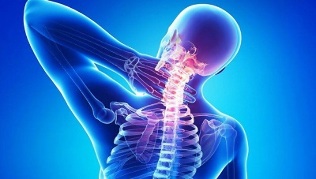
Due to the stretching of the joint capsules, the intervertebral discs lose their ability to fix the vertebrae. Such instability in the structure of the spine increases the risk of pinching nerve roots or constriction of blood vessels. This characteristic is characteristic of, for example, cervical osteochondrosis, which is accompanied by intense verbal symptoms.
Causes of the disease
The condition of intervertebral discs may worsen if the tone of the skeletal muscles of the spine decreases. Due to the irrational and asymmetrical work of the muscles, cartilage can be destroyed by permanently preserving the non-physiological position of the body. This violation is a consequence of wearing heavy bags on the same shoulder, using soft mattresses and high pillows.
The process of destruction of intervertebral discs is accelerated by a number of negative external and internal factors. These are:
- disorders of the endocrine mechanism and metabolic disorders; infectious
- pathologies, including in chronic form;
- spinal cord injuries in the form of compression fractures, bruises;
- regular and prolonged hypothermia of the body;
- systemic and degenerative-dystrophic diseases - gout, psoriasis, rheumatoid arthritis, osteoporosis, osteoarthritis;
- smoking and alcohol consumption, which disturbs the condition of the vascular system, impairs blood circulation and provokes a lack of nutrients in the cartilage;
- Insufficient physical development, posture problems, flat legs - these defects increase the load on the spine as there will be insufficient cushioning;
- obesity;
- genetic predisposition;
- Exposure to regular stress.
Symptoms
The main clinical sign of osteochondrosis of any localization (neck, chest or lumbosacral) is pain syndrome. With relapse, the pain penetrates, radiating to nearby areas of the body. It also grows with a slight movement. This forces the patient to place the torso in a forced position to minimize discomfort and pain:
- In cervical osteochondrosis, it will be preferable to turn the whole body instead of one head;
- when the chest form of the disease is present, it is difficult for the patient to take a deep breath, so he tries to minimize the depth and frequency of breathing to exclude acute chest pain;
- Patients with lumbar type disease have difficulty sitting down, moving upright, moving because the nerve at the site of the spine is constricted.
Patients usually complain of dull, persistent pain and stiffness in the morning after waking up. In this case, a differential diagnosis will be needed to help eliminate the risk of developing inflammation of the skeletal spinal muscles or myositis due to osteoarthritis.
Painful and depressive pains occur due to the compensatory tension of the muscle tissues. This condition is necessary to stabilize the range of motion of the spine. Persistent mild to moderate pain may occur with significant elongation of the intervertebral disc and may result from aseptic inflammatory changes.
Osteochondrosis of a separate localization is characterized by special symptoms:
- In osteochondrosis of the neck, there is pain in the neck area, in the upper limbs. Headache and numbness of the fingers are observed. If the disease is severe, the vertebral artery can become pinched. In this case, the patient begins to complain of a significant deterioration in health.
- Chest localization manifests itself in acute and painful back pain, visceral pain syndrome occurs in the heart region, right hypochondria, and abdomen. Patients complain of numbness, skin paraesthesia, shortness of breath, crunching of the vertebrae.
- Patients with lumbar osteochondrosis complain of increased back and lower limb pain during exercise. They are often diagnosed with disorders of the organs of the urogenital system, problems with male potency, dysfunctional ovarian disorders. During remission, the pain may decrease. However, the effect of the provocative factor leads to its renewal.
- If mixed osteochondrosis occurs, symptoms may occur in more than one zone at a time. This condition is characterized by a more severe course of the disease.
It should be borne in mind that the movement of vertebrae and the formation of osteophytes cause compression of the vertebral artery. It nourishes the brain and provides its cells with an oxygen component. When pressed, food is restricted, so the patient has problems with coordination, headaches, tinnitus, and arterial hypertension.
Consequences without treatment
The complicated course of osteochondrosis is due to the relatively rapid development of hernia in the intervertebral discs. Their appearance is accompanied by a posterior displacement of the vertebral structure. This provokes the rupture of the longitudinal type posterior band, which results in instability of the position of the disc, protrusion of some sections into the region of the spinal canal. Hernia rupture occurs when the core pulsed disc penetrates the area of the canal.
When abnormal abnormalities in the structures of the vertebrae appear, the back of the brain begins to compress, and the patient develops disogenic myelopathy. Symptoms of this condition are accompanied by numbness and weakness in certain muscle groups in the upper and lower limbs. Paresis, muscle atrophy and tendon reflexes are manifested. In some cases, there are problems with emptying the bladder and intestines.
Herniated discs are dangerous if they compress the arteries that supply the spinal cord. The result of this pathology is the formation of ischemic zones where neurons have undergone damage and death. Manifestation of the neurological effect is manifested in motor dysfunction, decreased palpation, and trophic abnormality.
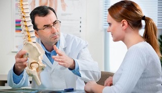
Disease Diagnostics
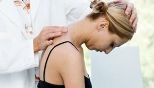
The initial diagnosis is based on the patient's complaints and symptoms. The person skilled in the art studies the condition of the spine in different positions, suggesting that the patient is at rest or in motion. In the next stage, the patient is referred to laboratory diagnostics to help clarify or refute the diagnosis.
The research methods used are as follows:
- Radiography- provides a complete examination of the spinal column by evaluating the condition of the vertebrae, the existing abnormalities in the form of growths and curvatures. The skilled artisan will be able to determine the intervals of the intervertebral type, the condition of the holes. A two-step X-ray is performed to accurately identify osteochondrosis localized in the chest or neck area. In the first stage, the patient lies on his side, and in the second, he lies directly on his back.
- Tomography using MRI or CTprovides highly informative data to help examine vertebrae in detail without interfering with the organs that cover them. The image shows the nerves and the vascular system. MRI helps identify signs of many diseases of the spine and the location of the damage. With CT, the hernias are visualized to determine possible deviations in the structure of the spine.
- Laboratory testto assess blood status and main parameters. It makes it possible to clarify the diagnosis and determine the possibility of the development of concomitant diseases.
In many cases, tests result in doctors diagnosing certain background conditions that are potentially dangerous for their complications. For example, we talk about hernia, protrusion, radiculitis. Proper diagnosis of problems helps to effectively treat osteochondrosis. In this case, the disease itself is disguised in the early stages of development as a symptom of other diseases.
Therapeutic process
Osteochondrosis is treated conservatively or by surgery. The choice depends on the severity of the condition, neglect, the level of tissue damage, and the reasons for its occurrence.
It is important to note that a complete cure for osteochondrosis is not possible because there are no medications to help completely recover discs and vertebrae. The therapeutic effect focuses on inhibiting the destructive process and increasing the duration and stability of remission.
Symptomatic therapy uses chondroprotectors based on chondroitin sulfate or glucosamine.
The efficacy of the therapeutic process using chondroprotectors has been clinically confirmed based on long-term tests. If you take these foundations for a long time from 3 months, there is a partial restoration of cartilage and other elements of the connecting type - the ligament-tendon device, the bursa.
Accumulation of glucosamine and chondroitin in the intervertebral disc area leads to analgesic, anti-edematous and anti-inflammatory effects. Therefore, there is a real possibility to optimize the dosage of NSAIDs, drugs belonging to the glucocorticosteroid group, muscle relaxants. You can expect a reduction in the patient's workload.
The efficiency of chondroprotectors is determined by the regularity of the input. Otherwise there will be no result. Ineffectiveness is also recorded in the treatment of grade 3 osteochondrosis, which is accompanied by significant cartilage destruction.
The following groups of drugs can be used to relieve pain:
- Non-steroidal anti-inflammatory drugshelps eliminate inflammatory disorders of soft tissues caused by dislocation of the vertebrae. NSAIDs are effective in reducing pain, swelling and stiffness.
- Glucocorticosteroid group devices- blockades are usually used in combination with an anesthetic. They are able to relieve pain, restore the immune mechanism and exert an anti-exudative effect.
- Muscle relaxers.They effectively fight muscle cramps due to nerve entrapment. They help relax skeletal muscles and block polysynaptic spine-type reflexes with an antispasmodic effect.
- External devices with a warming effect.Irritation of receptors in the subcutaneous tissue by activating blood flow is provided by special gels and ointments. These drugs have analgesic and anti-edematous effects.
Medical devices that activate blood flow can eliminate vertebrogenic-type symptoms that result from the localization of pathology in the neck or chest zone. Nootropics and drugs are also prescribed to improve microcirculation. In some cases, you may need to take antidepressants as well as anticonvulsants.
Physiotherapy is also used to treat osteochondrosis. UHF therapy, magnetotherapy, laser therapy, reflexology, massage, exercise, hirudotherapy, and swimming and yoga procedures may be prescribed. If conservative treatment is ineffective, the operation is performed by microdisectomy, defect disc valorization, laser reconstruction, or implant replacement.